Spatial Biology
Redefining tissue imaging with ultrafast, label-free QCL technology and IR-Guided MALDI.
Spatial Biology is transformed by the power of QCL (Quantum Cascade Laser) based Infrared Laser Imaging Microscopy (ILIM). Unlike traditional FTIR, ILIM offers unmatched speed, up to 180x faster, allowing for the high-definition chemical imaging of whole tissue sections in minutes. This label-free "molecular fingerprinting" serves as the perfect guide for downstream deep omics, such as MALDI Mass Spectrometry Imaging (MSI), enabling the "TissuePlus" workflow for targeted high-throughput analysis.
Our Solutions
High-Speed QCL Imaging
Leverage the brightness of Quantum Cascade Lasers to image large tissue areas at high spatial resolution (5 µm) in minutes, not hours.
- Scan whole mouse brains in ~7 minutes
- No cryogenic cooling required (Room Temp Detectors)
- Coherence reduction for artifact-free images
- Non-destructive and label-free
IR-Guided MALDI (TissuePlus)
Use the speed of ILIM to identify Regions of Interest (ROIs), such as tumor margins or specific cell types, to guide slow, high-cost MALDI analysis.
- Reduces MALDI acquisition time by >90%
- Target specific morphological subtypes (e.g., stroma vs acinar cells)
- Seamless ROI transfer to timsTOF fleX
- Correlate metabolic and proteomic data
Recommended Products
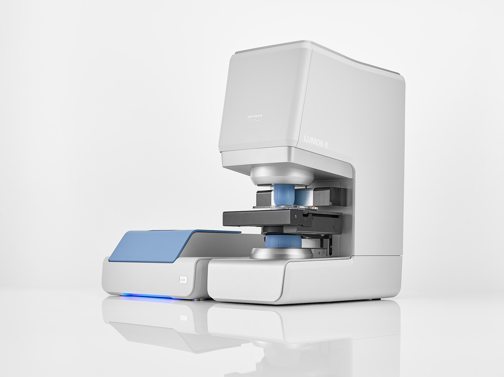
LUMOS II ILIM
The LUMOS II ILIM is a high-speed chemical imaging solution that combines powerful Quantum Cascade Laser (QCL) technology with a widefield IR camera to deliver rapid, artifact-free analysis of biological tissues, pharmaceutical tablets, and particles.
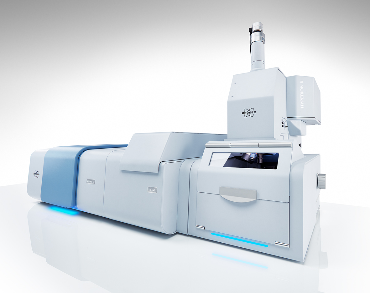
HYPERION II FT-IR & QCL Microscope
The HYPERION II is a premier research-grade microscope that uniquely integrates traditional FT-IR with high-speed QCL laser technology for advanced molecular imaging and sub-micron characterization.
Case Studies
Pancreatic Cancer (PDAC) Research
Bruker Application NoteDifferentiation of tumor subtypes and precursor lesions using ILIM-guided MALDI imaging.
Frequently Asked Questions
How is QCL/ILIM different from FTIR Imaging?
FTIR uses a thermal source and measures a broad spectrum, which is versatile but slower. QCL (Quantum Cascade Laser) uses a high-brightness tunable laser, allowing for discrete frequency imaging that is orders of magnitude faster (up to 180x) while maintaining high spatial resolution.
Can I use ILIM for live cell imaging?
ILIM is primarily designed for tissue sections and fixed samples. Its speed allows for high-throughput screening of large cohorts, making it ideal for pathology and pharmaceutical research rather than live cell dynamics.
What is the 'TissuePlus' workflow?
TissuePlus is a streamlined workflow where a sample is first rapidly imaged with QCL-IR (ILIM) to classify tissue types. These classifications are then used to automate the selection of measurement regions for downstream Mass Spectrometry Imaging (MALDI), optimizing both throughput and depth of coverage.