
LUMOS II ILIM: Spatial Biology Redefined
Imaging a tissue section chemically used to mean choosing between speed and information. A stain is fast and answers one question. A full infrared image answers many, and used to take hours.
The LUMOS II ILIM closes most of that gap. It uses a Quantum Cascade Laser instead of a thermal source, and Bruker states it delivers "up to 169x faster area scanning speed compared to FT-IR", at a spatial resolution of 4.25 micrometres.
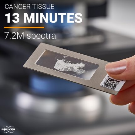
Unmatched Speed with QCL Technology
The LUMOS II ILIM utilizes a QCL source for Discrete Frequency Imaging. This enables whole tissue microtome section analysis in minutes. Traditional Globar-sourced FTIR imaging requires hours for similar analyses due to lower spectral power.
QCL-based imaging offers significantly higher spectral power at specific frequencies. This reduces acquisition times dramatically. The LUMOS II ILIM provides a high-throughput solution for spatial biology research.
Label-Free Spatial Biology: A New Paradigm
The LUMOS II ILIM provides non-destructive, stain-free analysis. It maps the intrinsic chemical architecture of tissues. This includes proteins, lipids, and metabolic markers, without the need for antibodies or dyes.
This label-free approach eliminates artifacts associated with staining procedures. It also preserves the integrity of the sample for further analysis. The LUMOS II delivers a more accurate representation of the tissue's chemical composition.
Clinical Relevance: Microtome Sections Made Easy
The LUMOS II ILIM is designed to handle microtome-cut tissue sections. This includes FFPE or cryosections, directly on standard IR-transparent slides. This streamlines the workflow for clinical pathology applications.
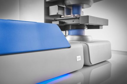
Sample preparation stays close to what a histology lab already does. Sections go onto IR-transparent slides in the standard format, so the step that usually blocks adoption of a new imaging method is not the sample handling.
Data Richness: Chemical Fingerprints for Deeper Insights
The LUMOS II ILIM provides detailed chemical fingerprints of tissue samples. These fingerprints offer deeper insights into tumor microenvironments. They also help with disease progression and cellular heterogeneity studies.
Metabolic profiling becomes accessible with the LUMOS II. Subtle chemical changes associated with disease states can be detected. This opens new avenues for biomarker discovery and diagnostic development.
Ease of Use: Accessible to All
The LUMOS II ILIM is designed for fully automated operation. This makes it accessible to biologists and pathologists. No specialized spectroscopy expertise is needed to operate the instrument.
The OPUS software runs the acquisition and the image processing. Bruker describes the system as having "full hardware automation with push-button software design", which is the difference between a shared instrument that gets used and one that waits for the single person who knows it.
Workflow Optimization with the LUMOS II ILIM
The route from block to image is short:
- Sample Preparation: Load microtome sections directly onto IR-transparent slides.
- Automated Acquisition: Set up the analysis parameters and initiate automated data collection.
- Data Analysis: Use OPUS software for spectral analysis and image processing.
- Result Interpretation: Extract chemical information and correlate it with histological features.
Technical Comparison: Globar vs. QCL
Traditional FTIR imaging with a Globar source delivers broad spectral coverage. However, it suffers from low spectral power, especially at longer wavelengths. This results in longer acquisition times and lower signal-to-noise ratios (S/N).
QCL-based imaging concentrates its power at selected frequencies instead of spreading it across the whole spectrum. Bruker states the result as "up to 169x faster area scanning speed compared to FT-IR" and imaging speeds "up to 62,400 spectra / second at maximum speed".
That speed has a price, and it is worth naming. Discrete frequency imaging measures the bands you selected, not the full spectrum. If you know which bands carry your answer, the trade is a good one. If you are still looking for the answer, a full FT-IR image on a smaller area remains the honest first step.
Where it fits, and where it does not
IR laser imaging is a chemical method, so it reports on proteins, lipids and other molecular structure. It does not identify a specific protein the way an antibody does. Groups that need molecular identity keep immunohistochemistry or mass spectrometry in the workflow and use infrared imaging to find the regions worth spending that time on. Bruker's IR-guided MALDI work is built on exactly that division of labour.
The spatial resolution is set by the wavelength of infrared light, so cellular detail is available and subcellular detail generally is not. For tissue architecture and regional chemistry that is enough. For anything smaller it is not, and no acquisition setting changes that.
See it on your own section
Speed figures from a datasheet say less than one of your own sections. I represent Bruker Optics in the Netherlands and Belgium, so I can arrange for a section of yours to be imaged on the system you are considering.
Arrange a measurement on your own tissue section and tell me which regions you care about and which configuration you are looking at.